
| Title | BPCC-102: IGNOU BAG Solved Assignment 2022-2023 |
| University | IGNOU |
| Degree | Bachelor Degree Programme |
| Course Code | BPCC-102 |
| Course Name | BIOPSYCHOLOGY |
| Programme Name | Bachelor of Arts (General) |
| Programme Code | BAG |
| Total Marks | 100 |
| Year | 2022-2023 |
| Language | English |
| Assignment Code | Asst /TMA / July 2022- January 2023 |
| Last Date for Submission of Assignment: | For June Examination: 31st April For December Examination: 30th September |

PART A-Assignment One
Answer the following questions in about 500 words each. Each question carries 20
marks.
1. Define hormone. Explain the structure and functioning of pituitary gland. Support
your answer with a suitable diagram.
Ans: Hormones are signaling molecules that are secreted by endocrine glands or cells, which travel through the bloodstream and act on specific target cells or organs to elicit a particular physiological response. Hormones play a crucial role in regulating various bodily functions, including growth and development, metabolism, reproduction, and stress response.
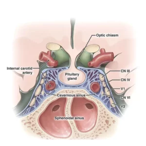
The pituitary gland, also known as the “master gland,” is a pea-sized gland located at the base of the brain, and it plays a central role in regulating the activity of many other endocrine glands in the body. The pituitary gland consists of two distinct parts: the anterior pituitary and the posterior pituitary.
The anterior pituitary is composed of specialized cells that secrete several different hormones, including growth hormone (GH), prolactin (PRL), follicle-stimulating hormone (FSH), luteinizing hormone (LH), thyroid-stimulating hormone (TSH), and adrenocorticotropic hormone (ACTH). These hormones are released in response to signals from the hypothalamus, which sends releasing or inhibiting hormones to the pituitary gland via the hypophyseal portal system.
The posterior pituitary, on the other hand, does not produce any hormones, but instead stores and releases two hormones produced by neurons in the hypothalamus: oxytocin and vasopressin (also known as antidiuretic hormone, or ADH). These hormones are synthesized in the hypothalamus and transported down the axons of neurons that terminate in the posterior pituitary. When the neurons are stimulated, the hormones are released into the bloodstream.
The structure of the pituitary gland is illustrated in the following diagram:
In summary, the pituitary gland plays a critical role in regulating various bodily functions, and it does so by secreting hormones in response to signals from the hypothalamus. The anterior pituitary secretes several different hormones that stimulate or inhibit the activity of other endocrine glands, while the posterior pituitary stores and releases hormones produced by neurons in the hypothalamus. The complex feedback loops that involve the hypothalamus and the pituitary gland help to maintain homeostasis in the body.
2. Describe the functioning of forebrain. Illustrate the lateral view of human brain.
Ans: The forebrain is the anterior-most part of the brain and is responsible for a variety of complex cognitive and behavioral functions. It consists of several important structures, including the cerebral cortex, thalamus, hypothalamus, and limbic system.
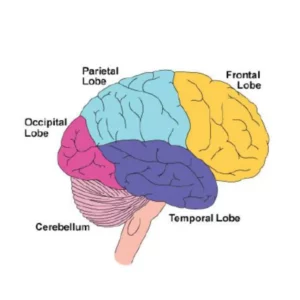
The cerebral cortex is the outermost layer of the forebrain and is composed of two hemispheres, each of which is further divided into four lobes: the frontal lobe, parietal lobe, temporal lobe, and occipital lobe. The cortex is responsible for a variety of higher-level functions, including perception, thought, memory, language, and consciousness.
The thalamus is a small, egg-shaped structure that acts as a relay station for sensory information from the body to the cortex. It receives sensory input from the eyes, ears, and other sensory organs, and then sends it to the appropriate area of the cortex for further processing.
The hypothalamus is a small, pea-sized structure located just below the thalamus. It plays a crucial role in regulating a variety of bodily functions, including temperature, hunger, thirst, and sexual behavior. It also controls the release of hormones from the pituitary gland and helps to coordinate the body’s response to stress.
The limbic system is a collection of structures that are involved in emotion, motivation, and memory. It includes the amygdala, hippocampus, and other structures that are located deep within the brain.
In addition to these structures, the forebrain also contains the basal ganglia, which are a group of structures that are involved in movement and coordination, and the corpus callosum, which is a thick band of nerve fibers that connects the two hemispheres of the brain.
The lateral view of the human brain is shown in the following diagram:
The cerebrum, which is the largest and most complex part of the brain, is located at the top of the brain and is divided into two hemispheres. The cerebellum, which is responsible for coordinating movement and balance, is located at the back of the brain, just below the cerebrum. The brainstem is the lower part of the brain that connects the brain to the spinal cord and is responsible for controlling a variety of automatic functions, including breathing, heart rate, and digestion.
Assignment Two
Answer the following questions in about 100 words each. Each question carries 5 marks.
3. Amnesia
Ans: Amnesia is a condition in which an individual experiences partial or complete loss of their ability to recall past events, memories, or information. The condition can result from various causes, including traumatic brain injury, stroke, Alzheimer’s disease, and substance abuse. There are two main types of amnesia: retrograde amnesia, which is characterized by the inability to remember events that occurred before the onset of amnesia, and anterograde amnesia, which is the inability to form new memories after the onset of amnesia. Treatment for amnesia involves addressing the underlying cause, as well as cognitive and behavioral therapies to help the individual cope with their memory loss.
4. Cranial Nerves
Ans: Cranial nerves are a set of 12 pairs of nerves that originate from the brainstem and exit through the skull. They are responsible for a wide range of sensory and motor functions, including controlling the muscles of the face, head, and neck, as well as transmitting sensory information from the eyes, ears, nose, and tongue.
The 12 pairs of cranial nerves are named and numbered as follows:
- Olfactory nerve (I) – responsible for the sense of smell
- Optic nerve (II) – responsible for vision
- Oculomotor nerve (III) – responsible for controlling most of the muscles that move the eye
- Trochlear nerve (IV) – responsible for controlling one of the muscles that move the eye
- Trigeminal nerve (V) – responsible for sensation in the face, as well as controlling the muscles of mastication (chewing)
- Abducens nerve (VI) – responsible for controlling one of the muscles that move the eye
- Facial nerve (VII) – responsible for controlling the muscles of facial expression, as well as transmitting taste sensation from the front of the tongue
- Vestibulocochlear nerve (VIII) – responsible for hearing and balance
- Glossopharyngeal nerve (IX) – responsible for transmitting taste sensation from the back of the tongue, as well as controlling some of the muscles of the throat
- Vagus nerve (X) – responsible for controlling many of the muscles of the throat and neck, as well as transmitting sensory information from the internal organs
- Accessory nerve (XI) – responsible for controlling the muscles of the neck and shoulders
- Hypoglossal nerve (XII) – responsible for controlling the muscles of the tongue.
These nerves can be tested for their function, and abnormalities in their function can help diagnose certain medical conditions.
5. Babinski Reflex
Ans: The Babinski reflex, also known as the plantar reflex, is a neurological reflex that is commonly tested during a physical exam to assess the function of the nervous system. The test involves stroking the sole of the foot with a blunt instrument, such as the handle of a reflex hammer, to elicit a response.
In a normal response, the toes will curl downward (plantar flexion) in response to the stimulation. However, in some cases, the big toe will move upward (dorsiflexion), and the other toes will splay out. This abnormal response is known as a positive Babinski sign or Babinski response, and it can indicate damage to the central nervous system.
The Babinski reflex is normally present in infants under the age of two, as their nervous system is still developing. In adults, a positive Babinski sign can be a sign of neurological conditions such as spinal cord injury, multiple sclerosis, stroke, or brain tumors. However, it can also be a normal variant in some individuals.
It is important to note that the Babinski reflex should only be performed by a trained healthcare professional, as improper or forceful testing can cause pain or injury.
6. Hemispheric Specialization
Ans: Hemispheric specialization, also known as cerebral lateralization, refers to the phenomenon where certain functions and cognitive processes are primarily controlled by one hemisphere of the brain, either the left or the right.
The left hemisphere of the brain is typically associated with language processing, mathematical reasoning, logical thinking, and analytical thought. It is often referred to as the “logical” or “analytical” hemisphere. The left hemisphere also controls the right side of the body.
The right hemisphere of the brain is associated with spatial awareness, creativity, intuition, and emotional processing. It is often referred to as the “creative” or “artistic” hemisphere. The right hemisphere also controls the left side of the body.
While it is clear that certain functions are primarily associated with one hemisphere or the other, it is important to note that the brain is a highly interconnected and dynamic system, and many processes involve both hemispheres working together. For example, language processing may primarily involve the left hemisphere, but the right hemisphere also plays a role in understanding figurative language and interpreting emotional cues in speech.
Studies have shown that some individuals have a stronger hemispheric dominance than others, and that this can affect their cognitive abilities and learning style. However, the extent and nature of hemispheric specialization can vary widely from person to person, and it is still an area of active research in the field of neuroscience.
7. The Z Lens
Ans: The Z lens, also known as the Nikon Z-mount lens, is a type of lens used for Nikon’s mirrorless cameras, including the Z50, Z5, Z6, and Z7 models. The Z mount is a relatively new lens mount that was introduced by Nikon in 2018 to replace its previous F mount, which was used for the company’s DSLR cameras.
The Z lens mount features a larger diameter and a shorter flange distance than the F mount, which allows for more flexibility and better performance in terms of image quality. The larger diameter also allows for faster autofocus speeds and better low-light performance, as well as the ability to create faster and more compact lenses.
The Z mount lenses come in a variety of focal lengths and apertures, including wide-angle, standard, telephoto, and macro lenses. Many of these lenses are designed with advanced features, such as fast and quiet autofocus, built-in image stabilization, and weather sealing to protect against dust and moisture.
One of the advantages of the Z mount is that it is designed to be compatible with Nikon’s F mount lenses through the use of an adapter. This allows users to continue using their existing lenses while gradually transitioning to the Z mount system.
8. Importance of Synapse
Ans: Synapses are the junctions between neurons that allow them to communicate with each other. They are an essential component of the nervous system and play a crucial role in many important functions, including learning, memory, and the control of movement.
The importance of synapses can be seen in the fact that the brain contains billions of them, each one playing a specific role in the transmission of information between neurons. When an electrical signal, or action potential, travels down the axon of a neuron, it reaches the synapse and triggers the release of neurotransmitters, which then bind to receptors on the adjacent neuron. This process allows the electrical signal to be transmitted from one neuron to the next, allowing for the rapid and precise communication between neurons that is necessary for many of the brain’s functions.
Synapses are also critical for learning and memory, as they allow for the strengthening and weakening of connections between neurons in response to experience. This process, known as synaptic plasticity, allows the brain to adapt and change in response to new information and experiences.
In addition, synapses play a crucial role in the control of movement. The brain sends signals through synapses to motor neurons, which then activate the muscles that are responsible for movement.
PART B- Tutorial
1. Explain the role of neurotransmitters in the effect of black widow spider venom.
Note: Diagrams or illustrations maybe used in support of your answer. Write the
answers in your own words. Please mention reference details of the articles/books, at
the end of your write-up.
Ans: Black widow spiders are known for their venom, which contains a potent neurotoxin called alpha-latrotoxin. This toxin acts on the nervous system by targeting the release of neurotransmitters, which are chemicals that allow nerve cells to communicate with each other.
Alpha-latrotoxin works by binding to specific receptors on nerve cells, causing an excessive release of neurotransmitters, particularly acetylcholine, which is involved in the transmission of nerve impulses to muscles. This overstimulation of nerve cells results in a variety of symptoms, including muscle pain, cramping, spasms, and paralysis.
The effect of black widow spider venom on neurotransmitters is similar to the action of some drugs, such as cholinesterase inhibitors, which are used to treat conditions such as myasthenia gravis and Alzheimer’s disease. However, the effect of the venom is much more potent and can cause severe, potentially life-threatening symptoms.
One of the most notable effects of black widow spider venom is the development of a condition called latrodectism, which is characterized by muscle pain and spasms, sweating, elevated blood pressure, and in severe cases, respiratory failure. Treatment typically involves the use of antivenom, which neutralizes the effects of the venom.
In summary, the venom of black widow spiders contains a neurotoxin called alpha-latrotoxin, which causes excessive release of neurotransmitters, particularly acetylcholine, leading to symptoms such as muscle pain, cramping, spasms, and paralysis. This effect is similar to the action of some drugs used to treat neurological conditions, but is much more potent and can be life-threatening. Treatment involves the use of antivenom to neutralize the effects of the venom.
Reference:
- Kuhn-Nentwig L, Stöcklin R, Nentwig W. Venom composition and strategies in spiders: is everything possible? Adv Insect Physiol. 2011;40:1-86. doi: 10.1016/B978-0-12-381387-9.00001-8.
- Binford GJ. Spider venom: from molecular structure to functional diversity and pharmacological potential. In: Annual Review of Entomology. Vol 57. 2012: 595-624. doi: 10.1146/annurev-ento-120710-100156.
2. ‘Older adult brains can generate new cells.’ Is this a myth or a reality? Support your
argument with case studies and evidence.
Note: Diagrams or illustrations maybe be used in support of your answer. Write the
answers in your own words. Please mention reference details of the articles/books, at
the end of your write-up.
Ans: The idea that older adult brains can generate new cells, known as neurogenesis, was once considered a myth. However, recent research has shown that neurogenesis can occur in certain regions of the adult brain, including the hippocampus, which is involved in memory and learning.
A landmark study by Peter Eriksson and colleagues published in 1998 provided the first evidence of neurogenesis in the human brain. The study examined postmortem brain tissue from adult humans and found that new neurons were being generated in the hippocampus. Since then, numerous studies have confirmed the presence of neurogenesis in the adult brain of several animal species, including humans.
In one study published in the journal Nature, researchers used a technique called carbon-14 dating to measure the age of human brain cells. They found that new neurons were being generated in the hippocampus well into old age, suggesting that neurogenesis is a lifelong process.
Other studies have shown that factors such as exercise and environmental enrichment can increase the rate of neurogenesis in the adult brain. For example, a study published in the journal Frontiers in Aging Neuroscience found that older adults who engaged in regular physical activity had higher levels of neurogenesis in the hippocampus compared to sedentary adults.
However, it is important to note that the extent of neurogenesis in the adult brain is still debated and may vary depending on factors such as age, health status, and environmental factors. Some studies have suggested that neurogenesis may decline with age or be impaired in certain neurological conditions, such as Alzheimer’s disease.
How to Download BPCC-102 Solved Assignment?
You can download it from the www.edukar.in, they have a big database for all the IGNOU solved assignments.
Is the BPCC-102 Solved Assignment Free?
Yes this is absolutely free to download the solved assignment from www.edukar.in
What is the last submission date for BPCC-102 Solved Assignment?
For June Examination: 31st April, For December Examination: 30th October